
I do not routinely perform bone grafting during All-On-X surgery.
While opinions on grafting during full arch surgery vary widely, in my experience, one of the more profound benefits of the AOX procedure is that it is often a graftless surgery.
In this week’s AOX Newsletter I discussed 4 reasons “Why I Don’t Routinely Bone Graft During AOX Surgery”.
While I am a firm believer that grafting is not often indicated in AOX surgery – there are times when I do feel bone grafting is advantageous.
Listed below are 6 scenarios in which I prefer to perform some degree of bone augmentation during AOX surgery.
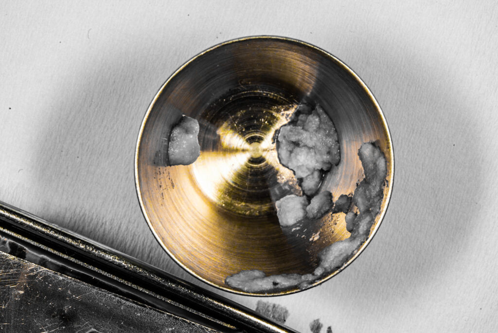
I should note that my preference of graft material is always particulate autogenous bone. This is normally bone debris taken during reduction (in the absence of active periodontal bone disease). A second, but less desirable choice for me, is particulate allograft. I also normally incorporate PRF into the graft and/or as a membrane.
6 Scenarios in Which I Prefer to Bone Graft During AOX Surgery
1. Grafting a buccal defect at an implant site.
I always want to avoid any type of buccal bone defect, and subsequent exposure of the facial implant threads, at an implant site.
Whether due to pathology, atrophy, or a buccal plate fracture – if there is any exposure of buccal implant threads, I always perform an only graft at this site and cover the graft with a PRF membrane (I do on occasion utilize a collagen membrane as well – this is case specific).
Not grafting in this scenario is asking for problems down the road with soft and/or hard tissue recession and defects at the implant site.
*It should be noted that my first preference in this scenario is to simply move the implant to an adjacent site and avoid the defect altogether. But sometimes this is simply not an option.
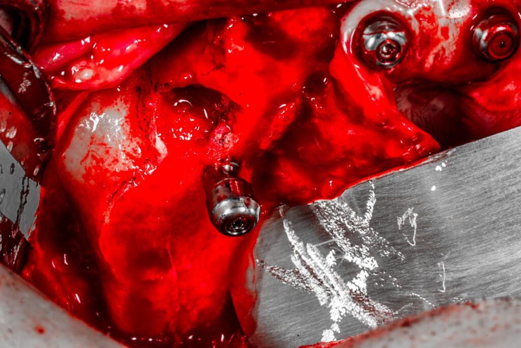
2. Grafting to assist with coverage of significant exposure of implant threads within a socket – as the implant traverses through the socket.
In this scenario, I am not referring to a buccal/lingual wall defect. However, depending on patient anatomy and the required reduction, an implant may pass through a socket with varying degrees of thread exposure within the bony housing of the socket walls.
There can also be loss of interseptal bone following removal of the dentition, essentially joining two adjacent sockets together. While the buccal/lingual walls are intact, the “open space” within the adjacent sockets can be notable.
For instance, placement of implant #4 may travel through the #4 socket as well as the #5 socket – with exposure of the threads inside the socket walls on either side of the existing or non-existent interseptal bone.
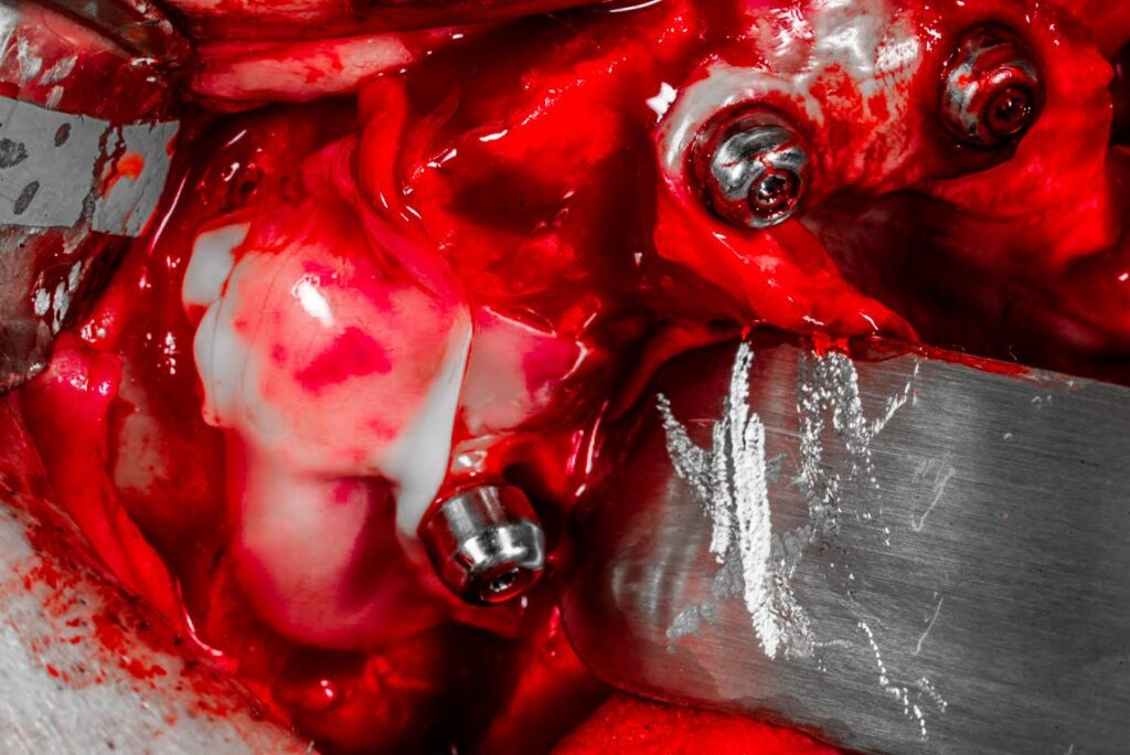
While this is not always the case, and or occurs to varying degrees – if I notice the implant threads to have significant exposure, I simply add a small amount of autogenous bone debris to those sockets. This assists with coverage of the implant threads. I also typically cover this area with a PRF membrane, overlaying the graft.
Does this make a difference? Honestly – I don’t know.
But it’s a very small graft with autogenous bone I already have. So, I go ahead and perform it.
This is not something I do every case – but I will perform this type of graft when I feel the intra-socket thread exposure warrants it.
3. Augmentation of a defect that remains following removal of an impacted maxillary third molar – if there is a possibility that a future pterygoid will be necessary.
I do not routinely graft impacted maxillary third molar sites. Nor do I even remove all impacted maxillary third molars during AOX surgery.
However, in exceptionally difficult cases that also present with impacted maxillary third molars – I think a little differently.
I attempt to always provide myself surgical outs. Therefore, in an otherwise challenging surgical case that also has impacted maxillary third molars, I will often remove these impacted teeth and consider a graft to the third molar sites. This is so that I have prepared the area for a pterygoid implant should the patient need one in the future (Or in reality, should I need one in the future to maintain the patient in a loaded prosthetic).
4. Grafting a defect that remains following removal of an impacted canine.
AOX cases with an impacted canine/canines can be challenging to say the least.
Often, the posterior and/or anterior implants will traverse through a portion of the impacted canine extraction site. For this reason, I normally graft these areas to provide full bony coverage of the implants in this region.
As discussed above, in order to leave myself surgical outs, I also normally perform a graft to encourage increased bone formation at the canine extraction site. Due to the size and location of these defects, augmentation helps create options and additional bone availability should an implant failure occur in the future.
5. Augmenting a large pathologic defect.
*Note that this augmentation occurs following either a pre-AOX biopsy and lesion confirmation, and/or if intra-operatively I am confident that the lesion is benign and adequately treated with enucleation.
It is important to know what pathologic lesion we are dealing with before grafting to ensure that the lesion has been, and is being, treated appropriately.
Following a proper diagnosis, large pathologic defects typically benefit from some type of grafting. This helps create future implant real estate, amongst other benefits.
Augmenting these defects also often helps provide better continuity and symmetry of the bone shelf.
Finally, a large pathologic defect adjacent implant sites tends to collect debris and be a hygiene nightmare – eventually leading to problems with the otherwise healthy neighboring implants.
6. A direct sinus lift utilized to provide an additional, future, posterior implant site.
I know it may not be popular or cool to post on Instagram, but occasionally I will still do an old fashioned sinus lift.
While I do not do this frequently, I do utilize this technique on occasion. For reference, I have performed direct sinus lifts on ~15 arches out of ~1,800 (assume half or more of those are maxillary arches).
I have typically utilized this technique when I am able to load the patient immediately, however, I felt that A-P spread was not adequate for long term implant health.
There are a plethora of surgical options I work through to improve A-P spread in these patients. However, if I cannot seem to find success with any of them, I will consider a direct sinus lift.
In this scenario, I perform a sinus lift on either the side of concern and/or bilaterally if indicated. I then load the patient (often with a shortened arch) but maintain their soft diet for 6 months. I return at the 6 month mark and add a posterior implant at the graft site and extend the prosthetic at this time, now with an eliminated cantilever.
Many people would combat this particular scenario with zygomatic implants. And that is totally fine. One could also argue benefits of zygomatic implants over sinus grafting. However, having been referred multiple zygomatic implant complications to manage from other practitioners – I’m not a huge fan of zygomatic implants. As such, I look for other solutions to these difficult cases. One of these options is the often overlooked sinus lift.
One of the most beneficial aspects of AOX surgery is its graftless nature. I embrace this attribute and do not often graft during my AOX cases.
That being said, I do feel the 6 scenarios listed above warrant at least a consideration for grafting, and can often have an improved outcome when bone augmentation is carried out.
Here’s to all the infrequent, but likely important times – we should, just maybe, consider ever so diligently – grafting a graftless procedure.
Matthew Krieger DMD
very nice article as always ,
what is your thought about leaving impacted canine if you can manage to place implant around it .
Hi Joseph,
I don’t think there is anything wrong with leaving an impacted canine IF you can adequately position your implants around it. That being said – I’ve done a number of these cases and never once have I encountered a scenario where the impacted canine was “out of the way”. It always seems to be somehow in the path of either the posterior implant real estate, the anterior implant real estate, or both.
Pingback: The 3 Most Common Ways I Utilize PRF in AOX Surgery - AOX Surgery
Pingback: Why I Don’t Routinely Bone Graft During AOX Surgery - AOX Surgery